
Specialist Advice — 7 minutes
When AI transform’s radiology
March 18, 2025

Radiology
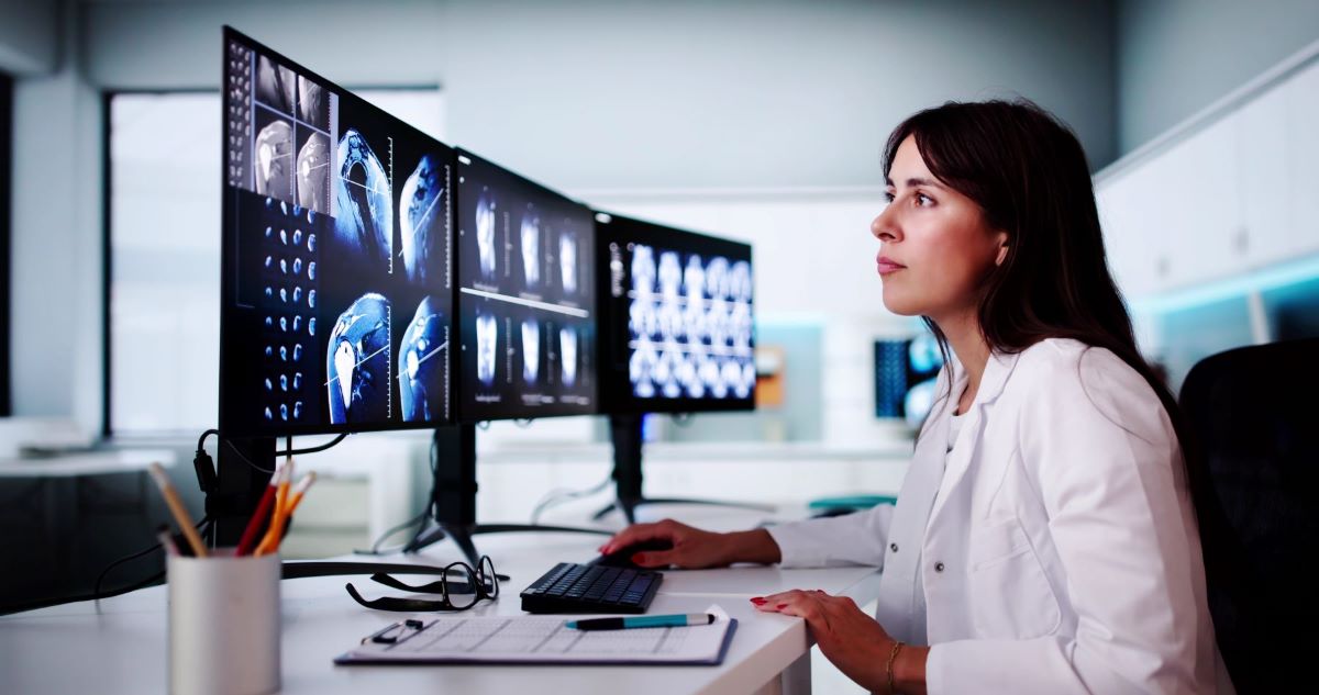
One of the primary medical technologies, radiology—x-rays, CT scans and magnetic resonance imaging (MRI)—play a central role in making diagnoses. Radiology produces 2- or 3-dimensional images which are mostly fixed but may sometimes also be dynamic. Just as pathologists use a microscope to analyze biopsies, radiological images need to be interpreted visually, one at a time, by radiologists. No matter how highly trained these specialists are, this is a painstaking verification, and the sensitivity of the analysis is limited by the eye’s ability to see the smallest details.
AI and image analysis
Over the past few years, there has been more and more talk about artificial intelligence. AI is a collection of techniques which can simulate our intelligence, to learn, reason, solve problems and make decisions. Since the beginnings of AI, one of its great strengths has been image analysis. As an example, facial recognition is now widely used to unlock our electronic devices, enter restricted sites or as a security tool for many governments and businesses worldwide.
How can AI recognize us in a crowd from a somewhat complete image of our face? It works by recording measurable facial characteristics, such as the distance between the pupils, shape of the ears or nose and skin colour in huge databases. Powerful computers can then compare our facial data with the data in their databases, almost instantly.
The database concept and the ability to make comparisons are two AI characteristics which can be harnessed in medicine. Another important strength of AI is its ability to learn to create new associations. For example, it may be able to establish connections between the growth of a lesion and characteristics such as density, homogeneity, border definition or distance between certain elements. Those parameters may vary from one lesion to another, but AI is able to perform a large-scale analysis and draw relevant correlations.
Radiomics
Like all images displayed on a screen, the images produced by radiological techniques are in two dimensions (think lung or bone x-ray photos) and can be broken down into pixels. If images are three dimensional (think CT scan), then we talk about voxels—the 3-dimensional equivalent of pixels. A scan section showing a tumour, for example, may contain millions of voxels. However, our eyes are not capable of analyzing complex combinations of these small groups of pixels. That is when AI becomes an invaluable analysis tool, since AI is able to interpret and derive information from them. [1]
This has led to radiomics, an approach which consists of extracting a very large number of characteristics and measurements from radiological images. The data are then analyzed by AI to build disease classification or prediction models.
Radiomics is currently showing the greatest potential in the field of diagnosing and monitoring cancers. Is the radiologist looking at an image with any significant abnormalities? If so, what type? In the context of monitoring cancer or suspicious lesions/abnormalities, are we seeing any changes from the previous exam? These are far from trivial questions. And yet, it is sometimes hard to find the answers with the naked eye. Thanks to AI, some cancers can be detected earlier. That leads to earlier treatment… but also brings a risk of overdiagnosis.
Early detection of breast cancer
A Norwegian study published in October 2024 showed that, in women who received a negative mammogram result, an AI-assisted analysis identified the women at higher risk of getting breast cancer in the next 4–6 years. The study compared the results of 3 mammograms performed every 2 years in over 116,000 women. Not only could AI easily identify abnormal results but also some subtle signs unnoticed during the initial analysis which were associated with a greater risk of developing breast cancer in the next 4–6 years [2]. Based on these results, we can well imagine that the women at highest risk will get enhanced monitoring!
Radiological images are interpreted one at a time by radiologists, in two stages:
- To identify suspicious images and set aside images which are unremarkable.
- To carry out a more detailed examination of suspicious x-rays.
AI can help radiologists tremendously with the first stage by sorting through the images, saving time for radiologists so they can concentrate on the more complex cases. In addition, since AI is able to extract information from a smaller number of images, that may shorten the analysis time.
Some radiology applications suggest that, for optimal quality, every image needs to be analyzed by two radiologists. However, it is hard to imagine applying this quality assurance policy to all x-rays produced across Quebec. In this context, AI could be considered as a backup observer, thereby improving workflow efficiency for radiologists.
Not only can AI help interpret images, but it can also help make the organization of work at radiology clinics and departments more efficient. For example, it can help manage the work of technologists and support staff. It can also support early detection of equipment-related problems.
AI’s limitations in radiology
Using AI in medicine raises multiple issues, including ethical ones [3].
AI relies on analyzing data from thousands of people. Many of those people might not be comfortable knowing that their medical data are being used without their knowledge for comparison purposes.
Another challenge with AI is its lack of explainability. Most of the time, we do not know what basis was used to build its decision-making algorithm. Not knowing may mask several issues, including the risk of discrimination against some minority populations such as Black or Indigenous people, who are often under-represented in the databases available.
This raises some key questions: who is responsible if there is an AI-generated mistake? The radiologists or the company that developed the software? And if AI is not rolled out uniformly from one clinic to another, might that accentuate social inequalities based on resources available?
In short, all these issues illustrate the limitations that must be considered when AI is used in medical settings.
In brief
Using artificial intelligence in medicine, including radiology, transforms health care by increasing the precision and rapidity of diagnoses. Even though there are some challenges, including ethics and understanding the models, it has clear short-term benefits: early detection, better management of complex cases and increased support for healthcare professionals. There is little doubt that radiomics, blending AI and radiology, is a promising innovation, provided we keep a healthy amount of vigilance.
Sources3
- R.J. Gillies, P.E. Kinahan and H. Hricak. Radiomics: Images Are More than Pictures, They Are Data. Radiology. February 2016, Vol. 278, No. 2, p. 563-577. DOI : 10.1148/radiol.2015151169. Accessed on March 28, 2025.
- J. Gjesvik, N. Moshina, C.I. Lee, et al. Artificial Intelligence Algorithm for Subclinical Breast Cancer Detection. JAMA Netw Open. 2024;7(10): e2437402. doi:10.1001/jamanetworkopen.2024.37402. Accessed on March 17, 2025.
- J.T. Martineau and F.R. Godin. Tour d’horizon des enjeux éthiques liés à l’IA en santé. Éthique publique. November 3, 2023 | 2023, http://journals.openedition.org/ethiquepublique/7978; DOI : https://doi.org/10.4000/ethiquepublique.7978. Accessed on March 17, 2025.