Everything you ever wanted to know about X-rays, CT scans, and MRIs
September 5, 2025

There have been major changes in medical imaging techniques since X-ray machines were first invented at the end of the 19th century [1]. Originally, these machines primarily made it possible to visualize bones and sometimes detect solid foreign objects.
The first CT scanners date back to the early 1970s. They can take a series of successive images and reconstruct the tissue in two or three dimensions. A few years later, a new type of scanner emerged, using magnetic resonance of the atoms in the tissue.
Each of these medical imaging techniques has its own unique characteristics: usefulness, accessibility, costs, as well as rare risks and discomforts. The myths surrounding each technique need to be debunked regularly.
X-rays
X-rays are electromagnetic waves, like light waves, but much more energetic. They can pass through solid objects, and we can use special devices to capture the waves going through the body. X-rays are easily stopped by dense items such as bones and microcalcifications, which appear bright white on images. Tissue, which is less dense, or soft tissue, partially blocks X-rays and appears as shaded areas with different densities. So, it’s hardly surprising that X-rays were first used for studying bones in order to diagnose fractures, bone deformations, bone cancers, osteoporosis, or dental cavities.
Even though soft tissue is not as obvious, some abnormalities can be detected. Examples are mammograms, chest X-rays (for diagnosing pneumonia or cancer), or bowel exams. Sometimes, injecting a radiopaque contrast agent helps distinguish specific abnormalities [2].
CT scan (computed tomography scan)
A conventional X-ray produces a 2D image. For a better view of a lesion’s outline or extent, X-rays need to be taken from a multitude of angles (from in front, from above, on an angle, etc.).
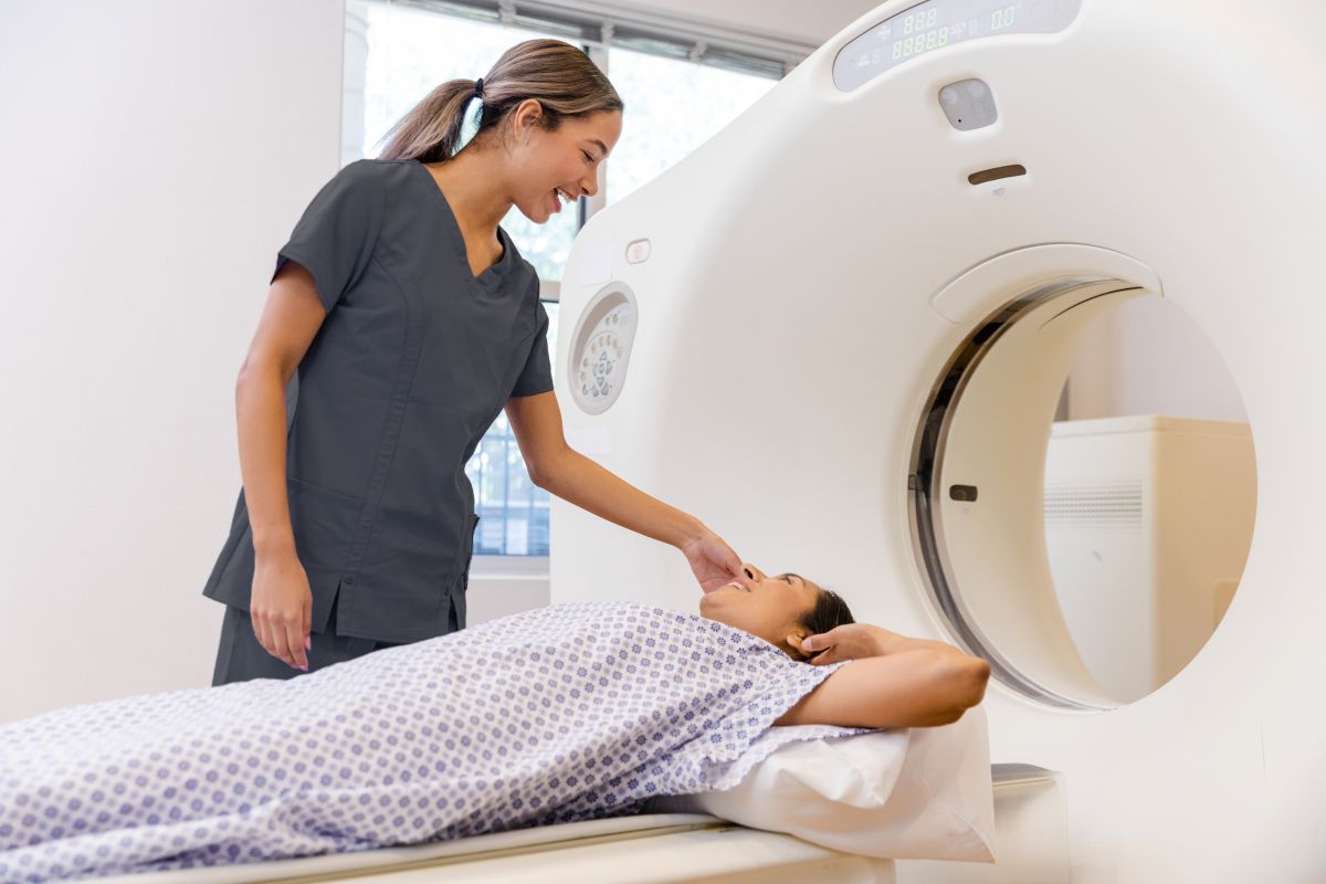
In the 1970s, the invention of CT scans made it possible to automate taking images from several angles, using an X-ray emitter rotating around the person [3]. Each rotation produces a large number of views, which are processed by software. The result: a precise 2D image of one complete slice of the body. Using a CT scanner, which looks like a large doughnut, we can analyze all parts of the body, from head to toe. The images obtained can also be combined to create 3D reconstructions. That means abnormalities’ shapes, extensions, and density variations can be assessed with greater precision.
Nowadays, CT scans are considered a second-line exam: they’re used when standard radiography or a single X-ray doesn’t provide enough information or details.
Radiation dose
X-rays are not completely harmless: at high doses or with prolonged exposure, they can cause adverse effects like burns, hair loss, or, less often, cell mutations, which may contribute to some cancers.
Luckily, modern machines have evolved considerably. The doses of radiation used for a standard X-ray nowadays are quite low. CT scans, which require numerous successive images, expose an individual to more radiation than a single X-ray does, but are still safe [4].
In practice, the risk of an X-ray is largely compensated for by the invaluable information that these exams provide for diagnosing. To limit unnecessary exposure, these tests are exclusively prescribed by a doctor and only when they are genuinely necessary. During pregnancy, although the risks to the fetus or children are low, special precautions are typically taken or imaging alternatives are used.
Magnetic resonance imaging
Magnetic resonance imaging (MRI) is different from X-rays and CT scans because it doesn’t use any radiation. Its action relies on a property of the nucleus of hydrogen atoms, very prevalent in water, and therefore in our bodies. In fact, water represents two thirds of our bodies’ total weight.
When someone is exposed to a very powerful magnetic field, the nuclei of hydrogen atoms align with the magnetic field but remain unchanged. If the aligned nuclei are bombarded with radio waves, the nuclei return to their original orientation and emit weak signals (resonance), which can be captured and transformed into an image [5].
Just like with X-rays, image quality can be improved by injecting a contrast agent, most often gadolinium [6].
MRIs can provide a more specific analysis of tissue content than CT scans can. This technology has become a fundamental tool for examining musculoskeletal, brain, abdominal, and pelvic lesions.
Metallic objects
Since MRI relies on a very powerful magnetic field, any metallic objects in the examining room will be strongly attracted by the machine. Metallic implants in the body such as pacemakers or metal fragments in the eyes may be moved within the MRI field. For this reason, they are contraindicated for an MRI. A questionnaire prior to the exam prevents any such situation arising.
Intense noises during an MRI
During an MRI, the machine makes a great deal of noise. Repeated banging sounds are often heard and may sometimes be as loud as a jackhammer. The noises are made by the coils creating the magnetic field.
To protect the ears, ear plugs or a headset will be offered. A headset allows technologists to communicate with the individual and even play music to lower anxiety [7].
Claustrophobia
During an MRI, much more of your body is inside the machine than when you have a CT scan. Being in that enclosed space may be particularly hard for anyone who has claustrophobia. Specific machines, called open MRIs, offer a larger opening than standard machines and may reduce the feeling of confinement.
CT scan or MRI?
A number of different imaging techniques may be used for making a medical diagnosis. How they’re different:
- cost (equipment, professional resources);
- risk (exposure to X-rays, contraindications related to pregnancy, metallic implants, and claustrophobia for an MRI, etc.);
- accessibility (no appointment needed for single X-rays versus a wait of several weeks for an MRI);
- and most of all their clinical usefulness.
There are also other diagnostic techniques, such as a clinical exam, ultrasound, endoscopy, and biopsy. They are typically used in succession, starting with simpler, more accessible methods and then, if necessary, using more complex exams.
Before prescribing an exam, healthcare providers assess clinical need and available technologies. No matter which diagnostic test is selected, it is always recommended that you discuss your questions and concerns about the exam with a healthcare provider.
Sources7
- H. Demirdjian. “La radiographie (I). Histoire de la découverte des rayons X et de leur application en médecine”. Culture SciencesChimie, October 2007. Accessible at https://culturesciences.chimie.ens.fr/thematiques/chimie-physique/la-radiographie-i-histoire-de-la-decouverte-des-rayons-x-et-de-leur. Accessed on September 4, 2025.
- M. A. Mafraji. “Radiographic Contrast Agents.” MSD Manual, April 2025. Accessible at https://www.msdmanuals.com/home/special-subjects/common-imaging-tests/radiographic-contrast-agents. Accessed on September 4, 2025.
- Let’s Talk Science. “Computed Tomography.” Accessible at https://letstalkscience.ca/educational-resources/backgrounders/computed-tomography. Accessed on September 4, 2025.
- Biron Health Group. “Myth to Fact – X-rays are a health hazard.” Accessible at https://www.biron.com/en/education-center/myth-or-fact/x-ray/. Accessed on September 4, 2025.
- Cité des sciences et de l’industrie. “De la tomographie au scanner.” Accessible at https://www.cite-sciences.fr/fr/au-programme/lieux-ressources/bibliotheque/ressources/ressources-en-ligne/dossiers/regards-sur-lhistoire-de-limagerie-medicale/de-la-tomographie-au-scanner#. Accessed on September 4, 2025.
- Ameli. “Comment se déroule une IRM?” February 2025. Accessible at https://www.ameli.fr/assure/sante/examen/imagerie-medicale/deroulement-irm. Accessed on March 5, 2025.
- Imagix, Biron Health Group. “Magnetic Resonance Imaging (MRI).” Accessible at https://imagix.biron.com/en/magnetic-resonance/. Accessed on September 5, 2025.
